Anatomy Between Hip Lower Ribcage In Back - Lower Back Pain Types Symptoms Treatment - Sechrest, md, narrates an animated tutorial on the anatomy of the hip joint.
Anatomy Between Hip Lower Ribcage In Back - Lower Back Pain Types Symptoms Treatment - Sechrest, md, narrates an animated tutorial on the anatomy of the hip joint.. Certain health conditions or injuries can affect the nerves in both of these areas. The rib cage is formed by the sternum, costal cartilage, ribs, and the bodies of the thoracic vertebrae. Structure of a typical rib the segmentation of the thorax is produced by both the intervertebral discs and the intercostal spaces between adjacent ribs. In this episode of eorthopodtv, orthopaedic surgeon, randale c. It includes over 30 bones, such as your femur and metatarsals, along with over 40 muscles, including your quadriceps and hamstrings.
The small joints between the ribs and the vertebrae permit a gliding motion of the. The ribs are curved, flat bones which form the majority of the thoracic cage. When pain in the lower back occurs alongside hip pain, there may be a common cause. Back rib pain or middle back pain is less common than lower back pain. The hip joint is a ball and socket joint that is the point of articulation between the head of the femur and the acetabulum of the pelvis.
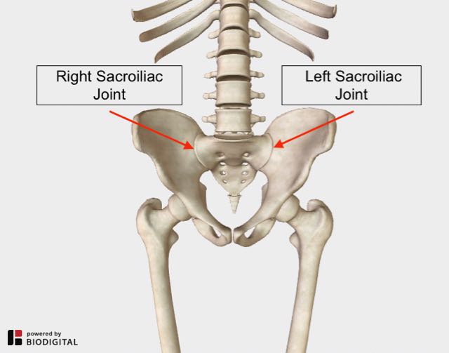
Lower Back And Hip Pain 7 Frequently Overlooked Causes from www.lower-back-pain-answers.com The skull, ribcage and pelvic bone are fairly solid and rigid parts of the body (though not always completely rigid). Sechrest, md, narrates an animated tutorial on the anatomy of the hip joint. The auricular surface articulates with the hip bones and is shaped like an ear. We hope you will use this picture in the. The vertebral column and thoracic cage. 1 hip anatomy, function and common problems. The space between the ribs is called the intercostal space. Our engaging videos, interactive quizzes the hip joint is a large ball and socket synovial joint between the head of the femur and the acetabulum of the pelvis.
It can help you understand our world more detailed and specific.
The hip joint is a ball and socket joint that is the point of articulation between the head of the femur and the acetabulum of the pelvis. During spinal flexion, the rib cage moves posteriorly, and the ribs are depressed. Your spleen also sits in the upper portion of the left side of your body, near your rib cage. The thoracic cage, commonly called the rib cage, provides protection for the 2 lungs, heart, esophagus, diaphragm and liver. Your lower extremity is everything from your hip to your toes, including your hip, thigh, knee, leg, ankle, foot, and toes. Anatomy is the amazing science. Sechrest, md, narrates an animated tutorial on the anatomy of the hip joint. Giving your body ample time to recover between activity sessions can reduce rib cage pain caused by damaged fascia. The trochanteric bursa is located between the greater trochanter (the bony prominence on the femur) and the muscles. It can help you understand our world more detailed and specific. Understanding lower back anatomy is key to understanding the root of lower back and hip pain. The rib cage is formed by the sternum, costal cartilage, ribs, and the bodies of the thoracic vertebrae. The human spine is composed of 4 sections of vertebrae.
We think this is the most useful anatomy picture that you need. Learn here what might cause rib cage pain, and the. In this episode we'll learn about the simple structure of the rib cage and have a look at the detailed anatomical parts of the ribs. Exhale and allow your rib cage and upper back come back to their natural position. You can click the image to magnify if you cannot see clearly.
Anatomy Of The Core Muscles Ankeny Ia Patch from patch.com Your rib cage provides a rigid framework for attachment of the muscles of your chest, shoulder girdle, back, diaphragm and upper abdomen. 1 hip anatomy, function and common problems. Back rib pain or middle back pain is less common than lower back pain. It can help you understand our world more detailed and specific. Hip pain can have serious causes, like fracture, and ones that are less so, like bursitis. They articulate with the vertebral column posteriorly. Sechrest, md, narrates an animated tutorial on the anatomy of the hip joint. We hope you will use this picture in the.
They articulate with the vertebral column posteriorly.
As they reach the side plane, they dive diagonally at about 45. These sections are cervical (neck), thoracic (upper and middle back), lumbar (lower back), and sacrum (tailbone). The costocorporeal joint is where the rib head connects with two adjacent vertebral bodies and the disc between them. The human spine is composed of 4 sections of vertebrae. Learn about the possibilities and when to see a doctor. Structure of a typical rib the segmentation of the thorax is produced by both the intervertebral discs and the intercostal spaces between adjacent ribs. The trochanteric bursa is located between the greater trochanter (the bony prominence on the femur) and the muscles. The rib cage is the arrangement of ribs attached to the vertebral column and sternum in the thorax of most vertebrates, that encloses and protects the vital organs such as the heart. Certain health conditions or injuries can affect the nerves in both of these areas. Pain coming from a person's rib cage may be nothing serious, or it may be a medical emergency, including a pulmonary embolism or heart attack. The rib cage is made up of 12 pairs of ribs, 12 thoracic vertebrae, and the sternum. From the back, the ribs angle down slightly. You can click the image to magnify if you cannot see clearly.
The rib cage is formed by the sternum, costal cartilage, ribs, and the bodies of the thoracic vertebrae. Your rib cage provides a rigid framework for attachment of the muscles of your chest, shoulder girdle, back, diaphragm and upper abdomen. The rib cage protects vital organs, such as the heart and lungs. Giving your body ample time to recover between activity sessions can reduce rib cage pain caused by damaged fascia. For example, a kidney stone can cause severe pain in the flank area (between the top of your hip and the bottom of your ribcage in your back).
Abdominal Muscles Location And Function from www.verywellfit.com Want to learn more about it? It is easy to overwork the lower back and hips because they are responsible for lifting, twisting, and moving the legs and trunk. Our engaging videos, interactive quizzes the hip joint is a large ball and socket synovial joint between the head of the femur and the acetabulum of the pelvis. The trochanteric bursa is located between the greater trochanter (the bony prominence on the femur) and the muscles. Your lower extremity is everything from your hip to your toes, including your hip, thigh, knee, leg, ankle, foot, and toes. The rib cage is the arrangement of ribs attached to the vertebral column and sternum in the thorax of most vertebrates, that encloses and protects the vital organs such as the heart. Structure of a typical rib the segmentation of the thorax is produced by both the intervertebral discs and the intercostal spaces between adjacent ribs. Anatomy is the amazing science.
You can click the image to magnify if you cannot see clearly.
1 hip anatomy, function and common problems. They're shapes that won't change the really useful thing is that from the front you can often see the lower front edges of it, it's centre line along the middle of the chest, and you know. Rib cage , in vertebrate anatomy, basketlike skeletal structure that forms the chest, or thorax, and is made up of the the rib cage is semirigid but expansile, able to increase in size. The costotransverse joint is where a small notch near the head of the rib (the tubercle) connects thoracic spine anatomy and upper back pain. We hope you will use this picture in the. Understanding lower back anatomy is key to understanding the root of lower back and hip pain. Learn here what might cause rib cage pain, and the. During spinal flexion, the rib cage moves posteriorly, and the ribs are depressed. The space between the ribs is called the intercostal space. They articulate with the vertebral column posteriorly. The hip joint connects the lower extremities with the axial skeleton. The tubercle is a bony prominence located at the junction between the neck and body which the costal angle also marks the attachment for some of the deep back muscles to the ribs. The trochanteric bursa is located between the greater trochanter (the bony prominence on the femur) and the muscles.
Share This :
Rhinokage Rio
Adalah seorang web designer yang suka mempelajari hal-hal yang baru seputar blog, template, coding dan Bisnis Online. Untuk mempelajari hal baru, membutuhkan kesabaran dan ketelitian dalam mempelajarinya.
Add Your Comments
Untuk menulis huruf bold silahkan gunakan atau .
Untuk menulis huruf italic silahkan gunakan atau .
Untuk menulis huruf underline silahkan gunakan .
Untuk menulis huruf strikethrought silahkan gunakan .
Untuk menulis kode HTML silahkan gunakan <code></code> atau <pre></pre> atau <pre><code></code></pre>, dan silahkan parse dulu kodenya pada kotak parser di bawah ini.
Halo Sobat Blanter, pada HUT RI ke-72 ini, ayo kita lebih semangat dalam mengejar cita-cita kita, untuk masa depan Indonesia yang lebih baik.Donasi yang kamu berikan akan saya gunakan untuk mengembangkan blog ini menjadi lebih baik. BANK BCA: 5475057811 a/n Sri Atmini PULSA : 0888-8905-441 (Smartfren) PAYPAL : paypal.me/blanter

:max_bytes(150000):strip_icc()/external-oblique-muscle-107702857-5bfd92bec9e77c002671fae7.jpg)

Tidak ada komentar:
Posting Komentar